Recent findings from a devoted team at Rice University have the potential to revolutionize our understanding of how cholesterol affects cellular membranes and their functioning, especially with implications for various diseases. Spearheaded by physicist and chemist Jason Hafner, the research has been documented in the esteemed Journal of Physical Chemistry. Cholesterol, a pivotal biomolecule, is integral to the composition of biomembranes—a sophisticated assembly of lipids and proteins that underpin cellular integrity. However, deciphering its nuanced behavior within these membranes has posed substantial challenges for researchers over the years.
One of the most crucial aspects highlighted by Hafner in this research is the influence of cholesterol on membrane organization, particularly regarding diseases such as cancer. Membrane structure is key to cellular function, and disrupting this organization can lead to severe pathological conditions. “Our breakthrough could have significant implications for understanding diseases related to cell membrane function, particularly cancer, where membrane organization is critical,” stated Hafner. This underscores the pressing need for research to grasp how cholesterol influences the microenvironment of cell membranes.
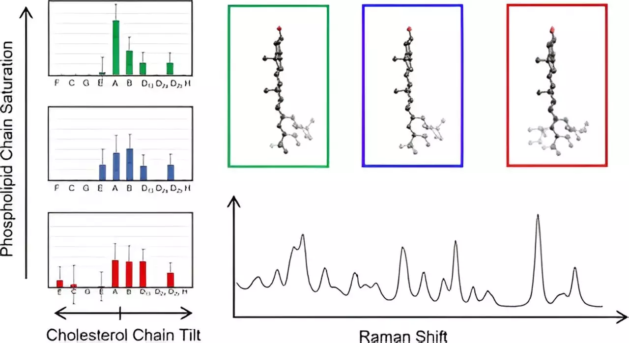
To navigate the complexities of cholesterol interactions within membranes, Hafner’s group employed Raman spectroscopy. This technique offers a sophisticated means of analyzing molecular vibrations using laser light, enabling scientists to delve deeper into molecular structures than previously possible. By examining cholesterol embedded within membranes, the researchers were able to correlate their experimental findings with theoretical predictions derived from density functional theory—an advanced computational methodology used in quantum mechanics. This blend of practical experimentation and computational analysis forms a robust framework for understanding cholesterol’s role with greater clarity.
Among the significant discoveries was the team’s identification of 60 different cholesterol configurations. They focused on analyzing the unique structure of cholesterol, characterized by its distinctive fused ring configuration and an eight-carbon tail. The study revealed intriguing patterns: cholesterol molecules were found to cluster based on how their tails deviated from the ring plane, indicating previously overlooked structural variations. This detailed examination not only sheds light on the diversity of cholesterol forms in membranes but also marks the first direct measurement of cholesterol chain structures in situ—within the membrane environment itself.
Surprisingly, Hafner and his team noted that within specific groups, all cholesterol molecules exhibited similar low-frequency spectra. This simplification allowed them to efficiently analyze and map out the various cholesterol chain structures within the membrane, enhancing our understanding of their organizational roles.
The ramifications of Hafner’s study extend far beyond mere academic curiosity. By unraveling the intricacies of cholesterol’s behavior within cell membranes, this research opens new avenues for exploring membrane-related diseases. This foundational knowledge could pave the way for targeted therapeutic strategies and deeper scientific inquiries into the pathology of diseases. As the team, including a diverse group of students and postdoctoral associates, continues to build on this research, the ultimate goal remains clear: to bridge the gap in our understanding of biomembrane dynamics and its broader implications for human health.

