In the rapidly evolving landscape of scientific inquiry, breakthroughs in imaging technologies are crucial to unlocking new frontiers. A collaborative effort spearheaded by Trinity College Dublin has achieved just that, unveiling a groundbreaking imaging method that significantly optimizes both time and radiation usage in electron microscopy. This cutting-edge development does not merely enhance existing techniques; it lays a foundational shift in how we understand and apply imaging in diverse fields such as materials science and medicine. The implications of such advancements are far-reaching, with the potential to radically improve our ability to visualize sensitive materials like biological tissues, which can be easily compromised during traditional imaging processes.
The Limits of Conventional Methods
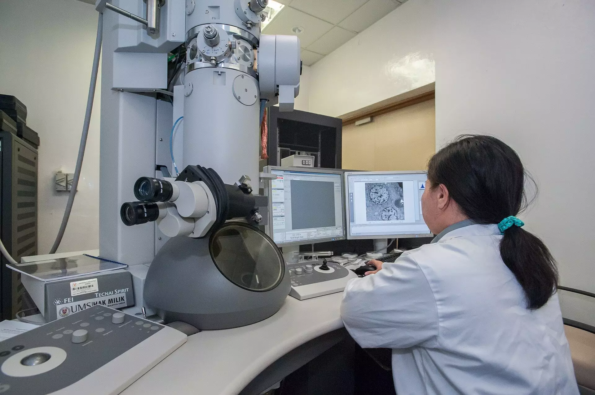
Traditional scanning transmission electron microscopes (STEMs) employ a well-established technique that involves meticulously directing a focused electron beam across a sample. This technique, while effective, is inherently limited by a standardized “dwell-time” for each point in the image. Just as a camera’s exposure time affects the quality of its photographs, the fixed duration during which electrons bombard a sample can lead to unnecessary strain and damage. The indiscriminate exposure across various regions of a sample generates an overwhelming amount of radiation that, although it contributes to the formation of an image, often at the expense of the sample’s integrity.
As scientists push the boundaries of what’s possible in imaging, the risks associated with excessive radiation exposure, particularly when dealing with delicate biological samples, have developed into a recognized challenge. In many instances, resulting images may not only be of poor quality but can also misrepresent the actual condition of the sample, further complicating research efforts.
A New Paradigm: The Event-Based Detection System
The breakthrough from Trinity College Dublin moves away from the conventional dwelling methodology to adopt an innovative event-based detection approach. Instead of relying on a fixed exposure time to collect signals from electrons scattered throughout the sample, this method intelligently calculates the time taken to observe a specific number of detected events. The ingenious pivot in thinking recognizes that not all electron impacts yield the same degree of useful information, a realization critical to reducing potential damage while maintaining image fidelity.
In essence, the researchers illustrate that the very first electron detected at a given location offers significant insights into the sample’s characteristics, while subsequent hits deliver diminishing returns in terms of information gain. Therefore, it becomes logical and advantageous to minimize the exposure of the sample to additional electrons. This increased efficiency allows for high-quality imaging that significantly lessens the radiation dose required to achieve results previously thought to necessitate a more intensive electron bombardment.
Introducing Tempo STEM: A Technological Leap
To bring this revolutionary concept into practice, the research team has patented technology branded Tempo STEM. By collaborating with IDES Ltd., they have developed a sophisticated “beam blanker” that modulates the illumination of the electron beam. This device can swiftly “shutter” the beam when the sample has reached an optimal imaging point, a feat that has never before been accomplished at such a rapid rate in response to real-time events.
Dr. Lewys Jones, one of the leading researchers, emphasized the significant advancement in microscope technology through this integration of existing systems, while showcasing the real-world application and benefit of such innovations. This leap not only facilitates obtaining high-quality images but also effectively protects the sample from unnecessary radiation. With radiation levels kept to a minimum, researchers can now feel more assured about the validity and reliability of the imaging data they collect, which is especially imperative in the study of complex biological systems.
The Bigger Picture: Implications Beyond the Microscope
The ramifications of this imaging breakthrough extend beyond just examining biological specimens or materials science. The insights gained could have transformative impacts in various domains, such as nanotechnology and semiconductor manufacturing, where imaging precision is crucial. Moreover, as the exploration of biomedicine continues to advance, refined imaging techniques can lead to greater accuracy in diagnostics, potentially fostering the development of tailored therapies. The ability to visualize delicate samples with minimal damage heralds a new era of scientific understanding — one that allows researchers to probe deeper, investigate intricately, and ultimately accelerate the quest for knowledge. The commitment to innovation and the resultant technological advancements mark a defining moment for the future of imaging, paving the way for more resilient and insightful scientific exploration.