A recent innovation from the University of California, Los Angeles, has introduced a transformative leap in 3D Quantitative Phase Imaging (QPI). The study, published in *Advanced Photonics*, highlights a wavelength-multiplexed diffractive optical processor that could redefine how we visualize and analyze transparent biological specimens and other weakly scattering materials. Conventional QPI methods often struggle with their dependence on multiple illumination angles and extensive computational resources. These limitations can be especially burdensome in high-stakes arenas like biomedical diagnostics, where timely results are crucial.
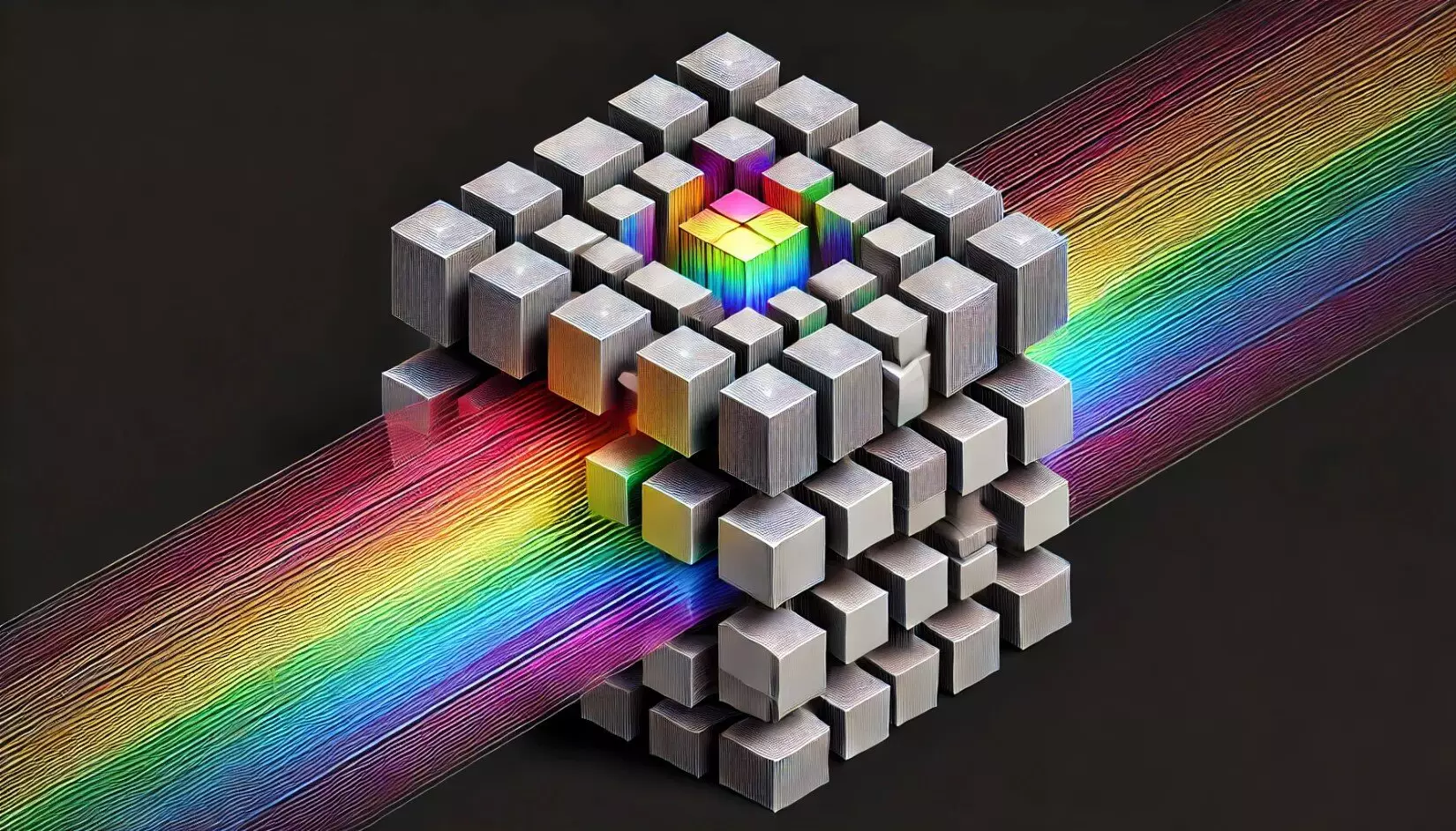
This new approach cleverly circumvents traditional bottlenecks by employing a system that simultaneously captures multiple 2D phase distributions at different depths, encoding them into distinct intensity patterns based on their unique wavelength channels. The beauty of this design lies in its capability to capture quantitative phase images without necessitating elaborate digital phase recovery processes. By relying solely on the intensity readings from a single image sensor, it significantly streamlines the imaging process.
Scientific Implications and Excitement
Aydogan Ozcan, the lead researcher behind this groundbreaking work, expressed profound excitement regarding the potential applications of this technology for biomedical imaging and diagnostics. The system promises a high-resolution, label-free imaging approach, which could drastically enhance our ability to monitor cellular and tissue dynamics in real time. Given that many of the currently used imaging techniques require the introduction of chemical labels or dyes—complicating the interpretation of the images and potentially interfering with the biological processes—the introduction of this novel processor stands to eliminate such confounding variables.
Moreover, this research collaborates the power of machine learning with optical design, utilizing deep learning algorithms to optimize the optical components involved in phase-to-intensity transformations. This synergy not only heightens the performance of the device but also enables rapid imaging of specimens across various axial levels, a significant advantage over traditional methods.
Versatility Across Spectrums
An exciting aspect of this new imaging system is its scalability and adaptability across various parts of the electromagnetic spectrum. By employing appropriate nanofabrication methods, the design could potentially be used in the visible range and infrared bands, expanding the horizons for various applications, from advanced laboratory research to practical field analysis. The potential integration with focal plane arrays further positions this technology as a frontrunner in the quest for efficient on-chip imaging and sensing solutions.
The broad implications of this study reverberate throughout many fields, including materials science, environmental monitoring, and, of course, medical imaging. By enhancing our capacity to analyze and diagnose diseases swiftly and accurately, it may even pave the way for earlier interventions in critical health conditions. This is crucial, as the quicker diagnoses can lead to better patient outcomes, especially in diseases where every second counts.
No longer constrained by technological limitations, the future of quantitative phase imaging appears not just promising but profoundly transformative. The implications of such breakthroughs may well extend past the realms of research and into everyday clinical settings, allowing healthcare professionals to deliver improved diagnostics and treatments that leverage real-time data visualization.