In the evolving landscape of neuroscience, understanding the intricacies of neural circuits is paramount for comprehending the brain’s functioning. Recently, the use of genetically encoded voltage indicators (GEVIs) has gained traction, as they offer a window into the electrical activity of neurons, shedding light on how information is processed within these vital networks.
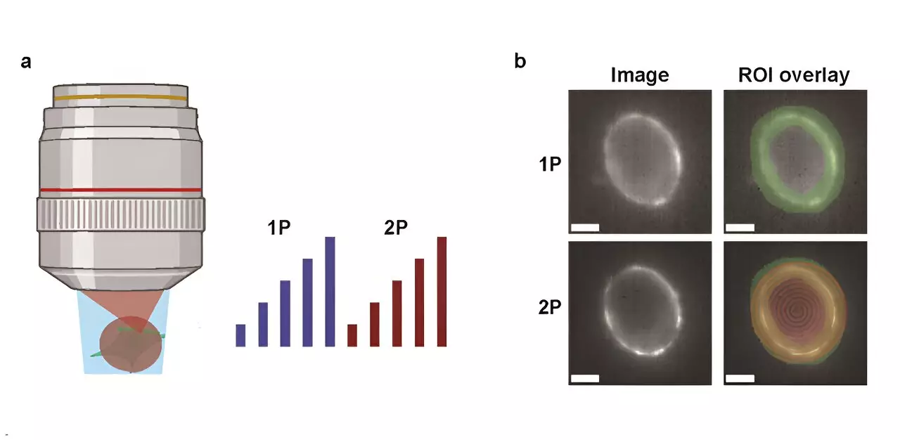
Among the methods employed for voltage imaging, the debate between one-photon (1P) and two-photon (2P) techniques remains contentious. A novel investigation conducted by Harvard University researchers provides a fresh perspective on this subject, delving into their respective advantages and limitations. By examining the specific optical and biophysical constraints paired with each imaging approach, the study contributes valuable data to this ongoing discussion.
The researchers conducted an in-depth analysis contrasting the brightness and voltage sensitivity of prevalent GEVIs, utilizing both 1P and 2P illumination. A critical component of their research focused on how fluorescence diminishes as penetration depth increases within the mouse brain—a vital consideration for in vivo studies.
Modeling for Understanding Neural Dynamics
To effectively interpret their findings, the research team developed a predictive model aimed at estimating the count of measurable neurons based on multiple factors, including reporter characteristics, imaging specifications, and the desired signal-to-noise ratio (SNR). This modeling approach not only aids in quantifying outcomes but also helps clarify the complexities involved in selecting the appropriate imaging technique.
A significant revelation from the study was the realization that achieving comparable photon count rates between 1P and 2P necessitates roughly 10,000 times more light intensity in 2P imaging per neuron. This formidable power requirement raises concerns about potential tissue damage and background noise, influencing the potential applications of 2P voltage imaging.
The implications of these findings are particularly noteworthy when considering in vivo scenarios. For example, when deploying the JEDI-2P indicator in the cortex of a mouse, researchers discovered that certain conditions—such as targeting an SNR of 10, limiting laser power to 200 mW, and operating at a frequency of 80 MHz—enabled the recording from approximately merely 12 neurons at depths greater than 300 µm. Such stringent constraints highlight the challenges of utilizing 2P imaging effectively for extensive neural population studies.
The current limitations associated with 2P voltage imaging—including insufficient voltage sensitivity and high photon-count demands—underscore an urgent need for advanced sensor technologies and imaging techniques. The insights derived from this research not only illuminate the critical trade-offs between 1P and 2P methods but also emphasize the importance of innovation in this field. As advancements continue to unfold, it will be intriguing to observe how future technologies evolve to enhance our understanding of neural circuits, ultimately enriching the domain of neuroscience.