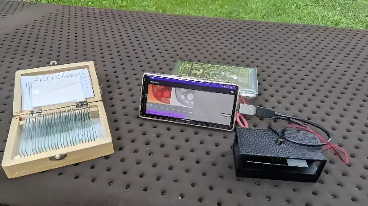
The advent of smartphones has significantly reshaped multiple fields, including healthcare, education, and scientific research. One particularly exciting innovation harnessing this technology is the smartphone-based digital holographic microscope, which offers high precision in 3D measurements. Researchers are optimistic about its potential due to its affordability, portability, and applicability in diverse settings, particularly in resource-poor environments.
Digital holographic microscopy (DHM) is an advanced imaging technique that reconstructs 3D information from captured holograms, revealing intricate details of a sample’s surface and internal structures. Traditional DHM systems are often encumbered by complex optical designs and the necessity for a personal computer to process holographic data, making them cumbersome for outdoor use or remote applications. This complexity limits their accessibility, particularly in educational and certain medical contexts.
The innovation discussed in this article addresses these limitations by simplifying the design and leveraging smartphone capabilities. The research team led by Yuki Nagahama from Tokyo University of Agriculture and Technology has developed a system that combines a straightforward optical configuration crafted through a 3D printer and relies on smartphone-based computation, rendering the system both affordable and transportable.
One standout feature of this new microscopic system is its capacity for near-real-time holographic reconstruction. By deploying a user-friendly smartphone interface, individuals can manipulate the display, such as zooming in on images using a pinch gesture on the touchscreen. This kind of interactivity enhances the user experience, making the process of examining biological samples or educational materials not only informative but also engaging.
The implications of such capability are far-reaching. In medical diagnostics, for instance, the low-cost design of this holographic microscope presents an opportunity for crucial applications, such as the diagnosis of diseases like sickle cell anemia in under-resourced regions. By providing access to sophisticated imaging tools, this innovation could help bridge gaps in healthcare delivery, enabling timely interventions.
Diving deeper into the technical foundation of this microscope, it functions by capturing the interference pattern produced by a reference light beam and the scattered light from the sample. This process generates a hologram, which is then digitally reconstructed to produce 3D information. However, past efforts in developing smartphone-based holographic systems faced challenges related to computing power and memory limitations inherent in mobile devices.
To overcome these challenges, Nagahama and his team implemented a novel technique called band-limited double-step Fresnel diffraction. This method optimizes data processing by minimizing the number of points calculated, thereby accelerating the holographic image reconstruction process. As a result, they achieved a frame rate of up to 1.92 frames per second under ideal conditions, demonstrating the microscope’s potential for near-instantaneous imaging of stationary objects.
In tandem with the technical advancements, the physical design of the microscope emphasizes portability. Utilizing lightweight materials and 3D printing technology to construct the optical housing, the team has created a device that can be easily transported and operated in a variety of environments. By incorporating a USB camera as part of the optical system and crafting a specialized application for Android devices, the researchers have ensured that users can operate the device with remarkable ease.
When the researchers tested their microscope with known patterns, they were able to successfully reproduce these images, confirming the accuracy of their design. Test cases included observing natural samples, such as the vascular structures within a cross-section of a pine needle, showcasing the robustness of the microscope’s imaging capabilities.
Looking forward, the research team plans to utilize artificial intelligence, particularly deep learning techniques, to refine and bolster the quality of the images produced. Digital holographic microscopy often encounters challenges such as the generation of unintended secondary images during hologram reconstruction. By investigating deep learning applications, the researchers aim to enhance the clarity and accuracy of the final reconstructed images while minimizing artifacts.
The smartphone-based digital holographic microscope represents a significant advancement in microscopy, offering an innovative solution for educational, medical, and research purposes. With its affordability, portability, and potential for real-time imaging, it paves the way for broader access to advanced diagnostic tools and groundbreaking scientific exploration. The future is indeed bright for this trailblazing technology.

