At first glance, the worlds of shimmering crystals and the diverse ecosystems inhabited by myriad species might seem worlds apart. However, a surprising relationship exists among various animals, such as fish, chameleons, and crabs, as they all engage in the fascinating process of crystal production. Unlike Walter White from the acclaimed series “Breaking Bad,” whose foray into the world of methamphetamine production was fraught with criminal intent, these creatures produce crystals for essential life-sustaining purposes—be it for communication, camouflage, or regulating temperature. The foundation of these diverse crystals rests upon two simple yet powerful molecules: guanine and hypoxanthine. The ability of different creatures to manipulate these basic ingredients into an array of structures raises questions about the biological strategies employed in crystal formation.
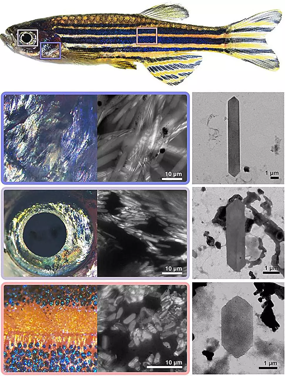
Central to the latest findings is the zebrafish, a vibrant freshwater species graced with a kaleidoscope of colors attributed to its internal crystalline structures. Research conducted at the Weizmann Institute of Science sheds light on the remarkable variety and complexity of these crystals. Upon close examination of different zebrafish tissues, researchers, led by Dr. Dvir Gur, uncovered a fascinating diversity: gill crystals exuded a silvery sheen, eyes reflected bluish hues, while skin crystals glimmered in vibrant yellows and blues. The acquired knowledge about these crystals—ranging in shape and size—deemed them ideal for research, allowing scientists to bypass the complications inherent in interspecies comparisons.
Using electron microscopy, the research team observed that variations in crystal morphology corresponded to the specific anatomical sites within the fish. Gills housed long, narrow crystals, while the eyes contained shorter forms. Such findings pointed to the influence of molecular ratios; specifically, the relative proportions of guanine and hypoxanthine significantly impacted crystal formation. This interplay of molecules evokes imagery akin to culinary art—similar to how a baker tweaks ingredient ratios to achieve desired textures and flavors in cakes or pastries.
By manually adjusting the ratios of guanine and hypoxanthine, scientists successfully replicated various types of zebrafish crystals in a laboratory setting, underscoring the intricate link between molecular balance and crystalline structure. However, guanine and hypoxanthine aren’t merely structural components for crystal creation; they play critical roles in DNA synthesis and overall cellular performance across the animal kingdom. Understanding how these molecules are regulated presents essential insight into the intricate processes that govern life.
To explore this regulation, Ph.D. student Rachel Deis focused on isolating iridophores, specialized cells responsible for crystal formation in zebrafish. Collaborating with a multidisciplinary team, they delved into the dynamics of how these cells generate and organize crystalline structures. The investigation revealed a two-sided narrative: iridophores possessed plentiful enzymes related to crystal building, yet exhibited a paucity of enzymes typically associated with similar functions in other cellular contexts.
This apparent contradiction prompted researchers to theorize that iridophores rely on a distinctive enzyme balance that governs the ratios of building blocks. This balance is critical; if disturbed, it could jeopardize both crystal integrity and cellular health. The iridophore enzyme landscape differed vastly between zebrafish and humans, with fish exhibiting multiple enzymes for guanine preparation. This genetic toolbox permits fish to craft unique crystal compositions while maintaining vital cellular processes.
The researchers sought to validate their theoretical insights through experimental manipulation. By engineering a zebrafish unable to produce the pnp4a enzyme—crucial for guanine crystallization—they observed significant changes in crystal morphology. The engineered fish exhibited fewer, square-shaped crystals in its eyes, mirroring skin crystals in design. This pivotal experiment reinforced the hypothesis that crystal properties are inherently linked to enzyme expression levels, confirming that even minor genetic alterations could lead to profound changes in crystal morphology.
The collaborative nature of this research emphasizes the power of interdisciplinary approaches in unraveling the intricacies of biological phenomena. By bringing together biologists, optical experts, and materials scientists, the team not only enhanced our understanding of animal biology but also drew inspiration from nature’s designs for potential applications in material science. This study exemplifies the beauty and elegance lying within the molecular simplicity of life, illustrating how fundamental building blocks come together to create complex structures that fulfill vital functions in the living world.
The study of crystal formation not only elevates our comprehension of biological systems but also opens a window into innovative materials inspired by nature’s designs. As we continue to study these phenomena, we are reminded of the intricate and often surprising connections that exist within the tapestry of life on Earth.

